Peri-implant soft and hard tissue management in the aesthetic zone

Introduction

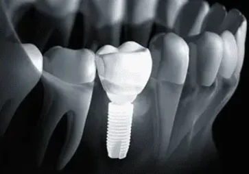
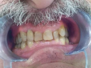
The survival of the dental implants is now predictable in almost every case but, the success of the implant still is a challenge according to its subjective basis. The aesthetic result is a criterion which is later achieved. It is good to know that to achieve aesthetics in the front area with an implant restoration is significantly more demanding than with the conventional restorations.

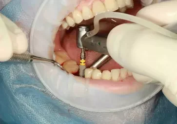
The insertion of dental implants in the front aesthetic area is a complex intervention which requires comprehensive planning and also a precise surgical procedure. Any other defects and complications later can lead to a disharmony with the perioral and facial structures. This could lead also to poor restorative and imperfect aesthetic outcome. The convincing point explains that dental implants which are inserted in a non-ideal position are exposed to non-axial loading. This situation can lead to high failure of the implant from a mechanical or aesthetic reason.

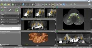
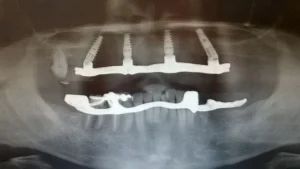
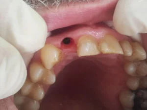
In order to achieve a stable and optimal function and also aesthetics, the implant position in the arch must be in a biologically tolerable and prosthetically driven location. One of the biggest challenges is the alveolar bone undergo specific changes after tooth extraction. It is a tooth dependent tissue and the tooth loss may lead to a loss in width and height of the alveolar process. This situation can cause shorter and narrower residual ridge which will later result with some complication with the ideal position of the dental implant. Sometimes, a soft and hard tissue management should be used.

What happens in the soft and hard tissue after a tooth removal?

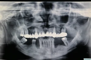
Patients should know that the periodontium is a complex of tissues which after a tooth extraction can be remodeled. This is explained with the fact that the alveolar ridge needs mechanical stimulation for maintaining its shape and density. After a tooth loss, there is a situation in which is noticed a decrease in trabeculation and also a loss of its width and height.

After a tooth removal, there are significant changes in the soft tissue. There is a noticeable loss of the gingival architecture which results in a reduction in the scalloped hard and soft tissue. The changes which occur involve the maturation of the wound. This process induces the calcification and the formation of the bone. The first phase is the clot formation which consists of a coagulum of white and red blood cells, inflammatory cells, and fibrin. After 4-5 days starts the second phase in which the coagulum is changed with a granulation tissue. Over a two week period begins the third phase in which the granulation tissue is changed with connective tissue. Following the fourth phase, starts the calcification of the osteoid of the socket base and periphery. The new formation made of bony trabeculae continues the next six weeks. The fifth phase is a complete epithelial closure if the wound. After 16 weeks, the socket is filled with bone and the osteogenic action is ceased.

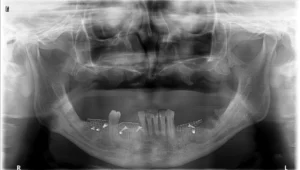
When it comes to the hard tissues after a tooth loss the alveolar bone undergoes the process of resorption. The dimensional changes are noticed in both vertical and buccolingual dimensions which result in a decreased gingival architecture, significantly manifested in the thin biotype. When macroscopically discussed, the healing procedure manifests in changes in the bone and also in the overlying soft tissues. There is noticeable bone resorption in a buccolingual part from 5-7 mm in the alveolar bone crest during 6-12 months. The reduction is more active in the first four months. The vertical dimension of the alveolar bone is also affected and there is a reduction of 2-4.5 mm. Another influence of the process over the bone resorption has an injury to the alveolus which can be occurred before or during the tooth removal intervention. It is marked as an iatrogenic fracture. It is possible that other local factors such as any infective process (periodontal or endodontic abscesses, tumors, or cysts) can contribute to a bone reduction.

How to make it perfect in the aesthetic zone?

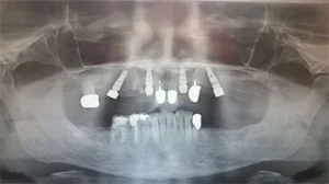
bone augmentationThe first thing an implant specialist often faces is the lack of sufficient bone in horizontal and vertical dimension. If this situation is not treated in the first stages of the intervention, it will definitely compromise the aesthetic and functional result. Studies have shown that many authors have approached the problem of the atrophy of the alveolar bone simply by suggesting combinations of surgical procedures and techniques in order to augment the defect. Because of this, many interventions, materials, and methodologies have evolved just to initiate the formation of the new tissue or to stop the further bone loss. Many grafting interventions have appeared utilizing autogenous bone grafts that were set as a gold standard for the process of bone augmentation.

The autogenous graft is a part of the patient’s bone and there are numerous advantages of its use. This type of graft consists of live osteoblasts and also osteoprogenitor cells that can proliferate and close the gap between the recipient bone and the graft. A successful outcome is marked because the microscopic architecture is perfectly fitted and also there is no immune reaction. These grafts result in the best regeneration of the bone defect because of the minimal postoperative resorption of the grafted bone. According to many studies, the preferred donor sites are grafts taken from the mandibular symphysis, followed by the ramus and the maxillary tuberosity.

It is good to know that there are also nonautogenous grafts and the most commonly used are demineralized freeze-dried bone. The bone formation features of these grafts are explained as osteoconductive and slightly osteoinductive. Keep in mind that this nonautogenous material can be utilized on a combination with autogenous bone grafts and also with resorbable or nonresorbable membranes.

Another type of nonautogenous material is the hydroxyapatite of b-tricalcium derivatives. Because this material when inserted alone does not have bone formation features, it needs to be utilized in a combination with some other autogenous or nonautogenous graft material.

Remember that the two mentioned nonautogenous graft materials are utilized for hard tissue augmentation.

The success of achieving great aesthetics around dental implants placed in the anterior part of the upper jaw has been a big challenge for the majority of dental clinicians. The key to an aesthetically satisfying appearance is the ability of the dental professional to properly manage the soft tissue around the dental implants. In some cases, there is no indication for soft tissue management.

In order to obtain long-term stability of the pink aesthetics around the dental implants, the dental professional may need additional techniques. This situation is in a strong correlation with the peri-implant soft tissue thickness which means a thick peri-implant biotype. If the dental professional diagnoses a thin biotype, then subepithelial connective tissue grafts or free gingival grafts may be used. The utilized grafts are taken for preventing the recession of the facial mucosal margin and also for permeation of the grey color gained from the dental implant. Keep in mind that the intervention of soft tissue augmentation can be done together with the implant insertion or during a second stage surgery intervention. According to literature, both technique alternatives have shown that can lead to a better aesthetic result and also can increase the thickness of the soft tissue.

The grafting process of the soft tissue can be used as a “rescue intervention” for managing the aesthetic complications which may appear with the implants. The usage of autogenous free gingival graft in the mucogingival interventions is very common. These grafts are considered as efficacious and reliable approach and they are most often utilized for increasing the amount of keratinized tissues around the dental implant. It is good to remember that free gingival grafts are still the gold standards for all situations where an increase in keratinized mucosa is needed. Usually, the donor site of an FGG (free gingival graft) is the highly keratinized hard palate. But, this way, the shade and the color of the recipient do not match with the adjacent soft tissues. Even though, FGG is used for increasing the keratinized tissue as a “rescue” procedure in order to cover the exposed implant threads.

tissue augmentation

Keep in mind that an FGG is utilized for those patients who have low smile lines, in cases where the color of the FGG would not compromise the aesthetic look of the implant site, and also when an extensive soft tissue augmentation intervention is desired.

Subepithelial connective tissue grafts are also utilized successfully for the management of the soft tissue defects and also for augmenting the contours of the alveolar ridge. Many of the procedures in which SCTG are used, can be performed directly tp the peri-implant soft tissue management and aesthetic optimization. In all cases, where they are properly utilized and are indicated, can provide significant gains and ensure stable condition in the soft tissue volume and contour. This will definitely contribute to a successful and the same time aesthetic management of the implant sites.

Summary

It is very important that all implantologists determine the present satiation of each patient individually. They should carefully consider the outcome of the surgical procedures and their timing in order to achieve a perfect and acceptable result. The implant specialist should notice the prime condition of the soft and the hard tissue architecture and later decide if hard or soft tissue augmentation procedures are necessary prior to the insertion of the dental implants. If the implant specialist confirms these interventions he should think which technique is the most appropriate. According to literature and the experience of the implantologists around the world, an autogenous graft material is a great treatment option for augmentation of the hard tissue especially when there is not a sufficient bone. Another important thing to consider is that the usages of block grafts which are taken from the mandibular symphysis are of the ramus are a great idea especially a large quantity of graft material is needed. In a proper relation to the timing for the implant insertion, the working protocol should follow the guidelines found in the literature and the final decision should be given for each patient on an individual basis.