Bone irregularities of the alveolar ridge and the jawbone can be acquired and congenital. Acquired irregularities can be result of:
- Individual and multiple teeth extractions performed in large time intervals, leading to uneven atrophy of the alveolar ridge;
- Not removed interdental or interradicular septum, after teeth extraction;
- Not made reposition of the walls of the alveolus, after tooth extraction;
- Fracture of the vestibular or oral cortical lamina as a result of applied inadequate technique of extraction;
- Surgical interventions of the alveolar ridge;
- Injury of the alveolar ridge, fracture of the jawbone, acute and chronic osteomyelitis.
Congenital alterations include torus palatinus and torus mandibullae.
Alveoloplasty
The bone irregularities of the alveolar ridge are corrected with a surgical procedure called alveoloplasty, which can be partial or total. Partial alveoloplasty is a leveling of a part of the alveolar ridge and it is more commonly applied. Total alveoloplasty is a leveling of the entire alveolar ridge and it is rarely applied. Alveoloplasty can be madeafter single or multiple tooth extraction and the technique of the intervention pretty much the same.
In conditions of missing teeth antagonists in one jaw, elongation of the alveolar ridge in the other jaw occurs, and the teeth get superposition. The elongation can be so much expressed that the teeth or the alveolar ridge of one jaw, can make a contact with the alveolar ridge of the other jaw. In this case it is practically impossible to make a prosthetic device, therefore, after the extraction of the tooth, we approach to the process of remodeling the alveolar ridge.
Technique: after the extraction of the tooth, a mucoperiosteal flap is made. When performing alveoloplasty, it is particularly important to maintain the depth (height) of the labial and buccal sulcus. This is achieved with making short incisions in the fornix and elevating small mucoperiosteal flap. We raise the flap and using bone plier we remove the serrated parts of the alveolus. Afterwards, we use a milling cutter to flatten the bone surface, but it is important to preserve the cortical lamina (as much as possible), which prevents occurrence of more pronounced resorption of the alveolar ridge, in the following period. The excess gingiva is cut off with scissors, the operative zone is washed abundantly with NaCl and the operative wound is closed with individual sutures.
Surgical correction of torus palatinus
When the torus palatines is discretely expressed (with a small dimension), it is not necessary to undertake surgical correction of the alteration, because in the preparation of the prosthesis, this area can be covered with foil on the working model. In this way the prosthesis does not directly press against it and it has good sability. If the torus is larger, it should be surgically removed because the prosthesis will be unstable and it may cause painful decubituses to appear. The best option in correction of palatinal torus is to do the intervention before pre-planned teeth extraction for making a total prosthesis. The presence of individual teeth, enables the preparation of palatinal plate, modified by a thermoplastic substance, which is applied in the patient’s mouth for a period of 7 to 10 days. It improves the comfort of the patient in the postoperative period, allows an intimate adaptation of the mucous flap to the bone tissue, prevents the emergence of pronounced postoperative hematoma, and a possible injury of the operative wound during mastication.
Technique: an initial incision is made along the middle palatinal line, with additional relaxation incisions at both ends. Such a designed incision prevents possible damage of the blood vessels (a. palatina major), provides adequate visualization and access, an operational field without tension. After rising the mucosa, it is fixed with traction sutures or with a wide periosteal elevator. If the present torus is small, the incision is made along the medial line with only one additional, anterior, relaxation incision (Y form). Such a torus can be removed using a large drill (with a pear-shaped form) or a milling cutter. When removing a larger torus, often lobulated, it is best to first make a groove along the center line, using a fissure drill. Then, the separation is made with additional, transverse sections through the torus. Further, the additional segments are removed with chisel or with bone pliers. The bone surface is flattened with a milling machine, the operative field is irrigated with NaCl and the wound is closed with individual sutures.
In totally toothless people, the incision is made along the alveolar ridge and the entire mucoperiosteal flap is lifted. Such a formed flap allows the setting of the sutures to be on a healthy bone base, away from the zone of removed palatinal torus.
Possible complications during the intervention: a fracture of the palatinal bone (when removing an unseparated large torus using a chisel), breakage of the thin palatinal mucosa and its consecutive necrosis (inadequate flap lifting), oronasal fistula (extremely rare complication).
Surgical correction of torus mandibullae
This bony protrusion, depending on its expression, can cause an injury of the mucous membrane during mastication, or lead to painful decubituses when the prosthesis is pressing against the mucous membrane of the torus.
Technique: After the mandibular anesthetic is given, we make an initial incision depending on the size of the torus and the presence or absence of teeth. In toothless patients the incision is made along the alveolar ridge and an additional, relaxation incision lingually, in front of the frontline of the torus. If teeth are present, then an envelope flap is performed around the necks of the teeth, and also we make a relaxation incision lingually, in order to achieve complete exposure of the bone exostosis. A piece of sterile gauze is placed under the torus, which prevents falling of small bone fragments between the tissues from the floor of the oral cavity, and the subsequent surgical procedure is identical to the one for torus palatinus.
Possible complications during this intervention are the breakage of the thin lingual mucosa and the possible occurrence of a postoperative hematoma.
Prosthetic fibrous hyperplasia of the mucosa
This condition is known as epulis fissuratum and it is most often localized in the vestibulum, in the area of the upper frontal teeth. Causes leading to this prosthetic hyperplasia are: trauma of badly made prosthesis, or in patients who have an upper total prosthesis and their natural teeth in the lower front. Namely, one or more folds are observed clinically, among which there are cuts in the mucosa that correspond to the prosthesis edges. The mucosa may be inflamed, in some places necrotic and painful on palpation and mastication. These hyperplastic creations reduce the retention and stabilization of the prosthesis, because they are mobile and in contact with the prosthetic edge. If epulis fissuratum is detected in the early stage when it is built from granulation tissue, removing the chronic irritation (non-usage of the prosthesis), may lead to its reduction or complete disappearance. In a later stage, when the granulation tissue has been transformed into fibrous, the condition is irreversible.
Surgical removal of the prosthetic hyperplasia is not performed at the first meeting with the patient, but the intervention is delayed for a period of 2 to 3 weeks, during which period the patient must not wear his prosthesis and must flash the oral cavity with oral antiseptic. During this period, the hyperplastic mucous membrane is reduced by 30% to 50%, which is much more convenient for surgical intervention compared to the initial condition.
Depending on the localization of the prosthetic hyperplasia, it is classified into 3 classes:
- I class – when it is located on the solid wall of vestibular fornix;
- II class – when it is located on the soft wall of vestibular fornix;
- III class – when it is located on the both walls, thus fulfilling the whole fornix.
Operational technique – I class
- After the application of local anesthesia, the epulis is fixed with surgical tweezers and then completely removed with a scalpel. In addition, the upper marginal part of the lesion are sutured with the exposed periosteum, so in this way, the height of the vestibular fornix is not reduced. The bare periosteum is covered with a prosthetic plate coated with zink-oxide impregnating paste, which the patient carries for the next 7 postoperative days.
Operational technique – II class
- The easiest class to remove. The epulis is removed with a scalpel and the lesion is directly sutured.
Operational technique – III class (for removing III-class lesion we use a combination of the two previous techniques)
Atrophy of the alveolar ridge and jawbone
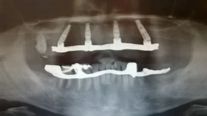
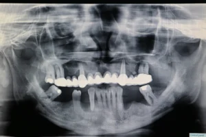
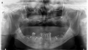
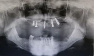
Loss of teeth and absence of physiological stimulations in the act of mastication, leads to atrophy of the alveolar ridge, which may be present along the entire ridge or only in individual parts. Deficient alveolar ridge may be compensated by implantation of a graft material, which can have a natural or artificial origin. Some of these materials also have an osteoinductive effect, they induce the growth of bone tissue through the porous material.
The technique of incorporating hydroxylapatite (alloplastic material) is relatively easy, and in some cases with two small vertical cuts, the entire alveolar ridge can be upgraded. In doing so, a subperiosteal tunnel is formed on the top of the alveolar ridge, which usually has a gully shape and it is suitable for application of the implant material using a specially designed syringe. The syringe is drawn in the tunnel and the area over the alveolar ridge is filled, starting from the most distant part of the tunnel to the vertical cut through which the syringe is drawn. In conditions of present extreme atrophy of the alveolar ridge, the only possibility is bone transplantation in combination with enosseal implants.
Surgical correction of the height of the alveolar ridge
The height of atrophic alveolar ridge can be surgically corrected in two ways: a relative increase and absolute increase. A relative increase of the height of the alveolar ridge is the usage of soft tissue surgery, most often in the vestibular fornix. An absolute increase is the usage of surgical bone transplantation procedures that increase or form a new alveolar ridge.
A relative increase in the height of the alveolar ridge is achieved by a procedure called vestibuloplasy. There are three techniques of vestibuloplasy:
- Deepening of the fornix with covering the two surfaces of the fornix with mucosa. From this group of operating procedures, the most commonly used is the submucosal vestibuloplasty (Obwegeser), which can be performed in the ambulance, with local anesthesia. The main indication for using this method is the presence of a healthy, flexible vestibular mucosa and a mild atrophy of the alveolar ridge. In doing so, we remove the excess submucosal tissue and do a reposition of the mucosa from the initial to a new (higher, deeper) level.
- Deepening of the fornix with secondary epithelization (two techniques)
– Deepening of fornix with secondary epithelization of the soft tissue surface of vestibular fornix (the surface of the alveolar ridge is covered with mucosa) – Kazanjian.
– Deepening of fornix with secondary epithelization on the surface of the alveolar ridge (the soft tissue area of the vestibular fornix is covered with mucosa) – Clark. - Deepening of the fornix by covering the operative lesion with a free graft (mucosa/skin). This operative technique is identical to the method of deepening the fornix with secondary epithelization of the soft tissue area of vestibular fornix – Kazanjian, with the difference that the operative lesion of the alveolar ridge is covered with a free mucous or skin graft. The basic indication for the usage of this method is partial deepening of the fornix, only on a certain part of the alveolar ridge.