Pre-prosthetic surgical interventions (correction of highly attached labial and lingual frenulum)

Successful mobile prosthetic rehabilitation in toothless patients is directly related to the presence of certain conditions, such as: sufficiently wide and high alveolar ridge, convenient correlation between the alveolar ridges, deep enough vestibular and lingual fornix etc. The absence of these conditions compromises the prosthetic rehabilitation, whereby it is necessary to perform pre-prosthetic surgical interventions in order to create conditions for making an aesthetic and functional prosthetic devices.

The minor pre-prosthetic surgical interventions are defined as corrective procedures of the soft-tissue and bony alterations in order to create conditions for successful prosthetic rehabilitation. They are usually performed under ambulatory conditions, using local infiltrative anesthesia.

All of these surgical interventions can be made preventively, in order to prevent the creation of irregularities that will obstruct the prosthesis of the patient, or correct the already present irregularities. In this case, atraumatic extraction of the teeth is particularly important. Therefore, rotational movements which protect the vestibular cortical lamina, and whose presence reduces the degree of subsequent atrophy of the jawbone, should more preferably be applied. During the extraction of the molars (especially in the upper jaw), we can separate the roots and surgically extract them, thus preventing a possible fracture of the vestibular cortical lamina and creating a bone defect or uneven alveolar ridge.

In general, irregularities that obstruct the making of a good and functional prosthesis are divided into two groups: developmental irregularities and gained irregularities. Developmental irregularities include: present mandibular and palatinal torus, low or high attachment of muscles, frenulums and plicae buccales, or atrophy/hypertrophy of the soft tissue. Acquired irregularities are usually result of teeth extraction, trauma and infection. The atrophy of the alveolar ridge is a significant problem. It happens as a result of its inactivity in toothless patients, who wear prosthesis. Atrophy of the ridge manifests as a non-favorable correlation between the movable and immobile mucosa, thus the quantum of the movable mucosa increases and it is inserted at the very top of the alveolar ridge. Also, injuries of the jaw bones and alveolar ridges, or surgical interventions on cysts or tumors, can turn into local factors that obstruct the making of prosthetic devices.

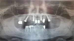
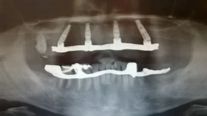
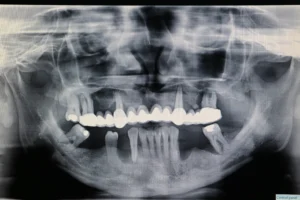
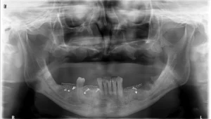
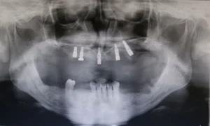
Before making a definitive prosthetic restoration, the condition of the alveolar ridge must be checked with clinical and radiological examination. On the basis of the obtained data, the intervention that is supposed to be correctly performed, is planned further. Otherwise, it is possible that while correcting one irregularity, we unexpectedly create another one (for example, poor partial alveoloplasy can create a shallow vestibular sulcus, a condition that adversely affects the stability of the prosthesis).

Normal anatomical correlations in the oral cavity may be impaired by congenital or acquired modifications in the soft tissue and bone segments of the alveolar ridge. One of them is the high attachment of frenulums.

Correction of the labial frenulum (frenoplasty)

The labial frenulum can be highly adhered (inserted) to the alveolar ridge, even at its very tip. This condition may be congenital or acquired as a result of a strongly expressed atrophy of the alveolar ridge. Under such conditions, the making of a functionally stable prosthetic device is difficult. Also, the labial frenulum can be removed because of orthodontic reasons. It may cause an appearance of diastema mediana in children, which is corrected by excision of the frenulim. A surgical removal of the frenulum is called frenoplasty and two surgical methods are most commonly used: excision frenoplasty and „ Z “ frenoplasty.

Excision frenoplasty

It is indicated under conditions where there is a residual alveolar ridge with a good vertical dimensions and a deep labial vestibulum.
Technique: after a given local anesthetic, the lip is pulled up, the frenulum is fixed with two Peon forceps, which are positioned at the upper and the lower end of the frenulum. An incision is made only through the mucosa, so that after removing the frenulum, the periosteum remains intact. When setting the sutures, in order not to reduce the labial fornix, the first suture is placed high in the vestibulum, which additionally reduces the postoperative hematoma.

„ Z “ frenoplasy

It is indicated when there is an alveolar ridge with a loss of vertical dimension and a shallow labial vestibulum.
Technique: elliptical incision is made, the fibrous frenulum if removed in a depth of several millimeters. Thereafter, two short incisions are made at the ends of the initial incision.

Correction of the lingual frenulum

The lingual frenulum can cause partial or complete ankiloglossia, which is a congenital anomaly. This condition is due to insertion of the frenulum to the floor of the oral cavity or to alveolar mucosa, and due to an extremely short frenulum that is connected to the top of the tongue. Ankiloglossia limits the movement of the tongue and thus creates problems with speaking, swallowing, mastication. Additionally, the lower total prosthesis moves from its place at each movement of the tongue.

Technique: after the application of a local anesthetic for n. lingualis, the tongue is immobilized and raised upwards. By raising the tongue, the frenulum is straining itself. Subsequently, convergent incisions are made on both sides of the frenulum, which interconnect at the ends. These incisions converge to the base of tongue. After removing the frenulum, the edges are flattened with scissors for obtuse dissection and the wound is sutured with individual sutures. The following anatomical structures can be damaged during the intervention: caruncula sublingualis, ductus sublingualis, ductus submandibularis, a. et v. sublingualis.